Visual field defects do more than tell us something is wrong. They tell us where it’s wrong. When the patterns of loss align with the anatomy of the visual pathway, clinicians gain a powerful shortcut to diagnosis. For optometrists and GPs, recognising these classic patterns transforms routine field data into actionable insights — guiding urgent referral, targeted investigations and early intervention.
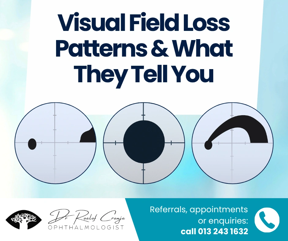
This article breaks down three foundation patterns: bitemporal, arcuate and central field loss — what each signifies, how they present, and why they matter. We also include a case snapshot of a seemingly routine exam that revealed a hidden pituitary lesion through pattern recognition alone.
Bitemporal Field Loss → Optic Chiasm
When the lateral (temporal) halves of both visual fields shrink or disappear, one should immediately think of the optic chiasm. The crossing fibres from the nasal retinas of each eye carry temporal field information; a chiasmal lesion disrupts them, producing a so-called bitemporal hemianopia. MSD Manuals+1

Key features:
- Loss of visual field on the outer halves of both eyes, respecting the vertical midline.
- Often gradual, subtle in early stages, and may show asymmetry.
- May occur without dramatic acuity loss or obvious fundus changes — making field testing crucial.
Common aetiologies:
- Pituitary adenoma (most common)
- Craniopharyngioma, meningioma, aneurysm near the chiasm EyeWiki
- Occasionally optic nerve tumour impacting the chiasm junction
- Early glaucoma can sometimes mimic with temporal wedges; always correlate with anatomy.
Clinical workflow tip:
Any bitemporal defect = neuro-imaging + urgent ophthalmology referral. For the GP or optometrist, this should trigger endocrine and neurosurgical liaison early, not just glaucoma work-up.
Arcuate & Nasal-Step Defects → Glaucomatous Pathway
Visual field loss in an arcuate (bow-shaped) pattern or a nasal step is emblematic of glaucomatous optic neuropathy. The retinal nerve fibre bundles arc out from the disc and respect the horizontal raphe — hence the familiar “arcuate” scotoma. MSD Manuals+1

Typical features:
- Paracentral or arcuate defect adjacent to blind-spot then arching nasally
- The defect “respects” the horizontal midline; seldom crosses it early
- A nasal step: difference in sensitivity between superior and inferior field often an early sign
Common causes:
- Primary open-angle glaucoma (POAG)
- Normal-tension glaucoma
- Secondary glaucomas (exfoliative, pigmentary)
- Severe high myopia may show similar patterns; always correlate with optic disc and OCT appearance.
Clinical workflow tip:
If you see an arcuate/nasal-step defect, ask: is the optic disc cupped? Are RNFL and GCC thinning present on OCT? Is IOP elevated? Field testing should be repeated for reliability and then glaucoma referral initiated early.
Central Field Loss → Macular or Optic-Nerve Focus
Central or centro-cecal defects (within the central 5°-10°) suggest pathology centred on the macula or papillomacular bundle of the optic nerve. Examples include dense central scotomas or subtle central/paracentral losses. American Academy of Ophthalmology+1

Important clues:
- Loss around fixation or a dense central defect
- Colour vision may be disproportionately affected (suggesting optic nerve involvement)
- Macula disease usually presents with central distortion or blind spot plus fundus/macular OCT changes
Aetiologies to consider:
- Macular: AMD, macular hole, CME, toxic maculopathy
- Optic nerve: optic neuritis, nutritional/toxic optic neuropathy, compressive or infiltrative optic neuropathy
- Early glaucoma can also affect central vision but typically later stage
Clinical workflow tip:
Central field loss → always examine the macula with OCT + fundus. If optic nerve involvement suspected, refer for neuro-ophthalmology and neuro-imaging promptly (especially bilateral, rapid onset or with pain on eye movement).
Integration in Clinical Practice
Step-by-step approach for GPs & optometrists:
- Review pattern: central vs arcuate vs bitemporal
- Check anatomical implications (pre-chiasm, chiasm, post-chiasm)
- Correlate with disc/OCT/I OP/fundus findings
- Decide urgency: glaucoma versus neurology referral
- Document, repeat repeatable fields, communicate findings clearly
Key reminder: Visual field defects are not just a number on the perimeter — they reveal underlying structural pathology. A field pattern that “matches” the anatomy can alert you to serious disease earlier than symptoms or basic exam findings.
Conclusion
Visual field testing remains one of the most under-utilised diagnostic tools in general practice and optometry. But when interpreted through the lens of anatomy — bitemporal for chiasm, arcuate for glaucoma, central for macula/nerve — field defects become powerful localising tools.
You don’t just measure vision — you interpret what the field tells you. This simply informed approach empowers GPs and optometrists to act earlier, refer smarter and ultimately improve outcomes.