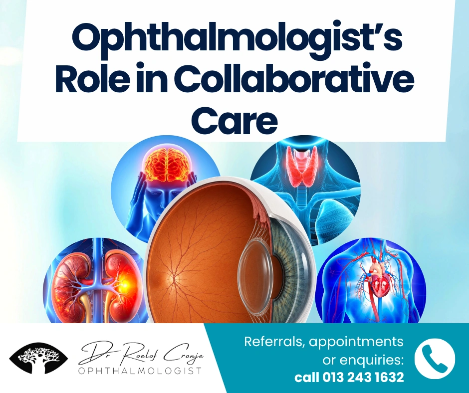
How collaborative practice improves outcomes for patients with complex, overlapping conditions
Modern medicine is rarely a solo act. Patients don’t arrive with neatly packaged “eye problems” or “systemic problems” — they arrive as whole people with interconnected conditions. Ophthalmologists are often the first specialists to spot signs of vascular disease, autoimmune activity, neurological dysfunction or metabolic imbalance.
This makes the ophthalmology clinic a vital entry point into multi-disciplinary care. With the right communication channels, many conditions can be diagnosed earlier, treated faster and managed more safely.
Below are hypothetical but realistic case stories that illustrate how collaborative care genuinely changes outcomes.
Case 1: The Silent Stroke Risk Identified in an Eye Exam
Ophthalmology + GP + Cardiology
A 62-year-old man presents with mild blurred vision. His fundoscopy shows a branch retinal artery occlusion (BRAO). He has no neurological symptoms and no history of stroke.
In the ophthalmology setting, sudden retinal ischemia is treated as a stroke equivalent — meaning the same systemic risk applies. After urgent communication with the GP, the patient undergoes:
- carotid Doppler ultrasound
- ECG and rhythm monitoring
- lipid and glucose screens
- initiation of antiplatelet therapy
Imaging reveals high-grade carotid stenosis. A vascular surgeon becomes involved, and the patient undergoes endarterectomy within a safe time window.
Outcome: Retinal damage is limited, but more importantly, a high-risk stroke pathway is prevented. The collaborative approach turns an eye finding into a life-saving intervention.
Case 2: Optic Nerve Swelling That Wasn’t Just an Eye Issue
Ophthalmology + Neurology + Emergency Medicine
A 26-year-old woman arrives with photophobia and painful eye movements. The optic disc is swollen, but the swelling is unilateral and accompanied by color desaturation and reduced visual acuity. These features suggest optic neuritis, not papilloedema.
An urgent call to neurology leads to:
- MRI brain and orbits
- Screening for demyelinating disease
- High-dose IV corticosteroid therapy
MRI shows white-matter lesions consistent with multiple sclerosis. Because the diagnosis is made early, she is started on disease-modifying treatment within weeks.
Outcome: Vision recovers well, long-term neurological decline is slowed, and the patient receives early counselling and support.
Without ophthalmology recognising the pattern, her diagnosis could have occurred months later — often after a more disabling relapse.
Case 3: The “Diabetic Eye Check” That Revealed Kidney Disease
Ophthalmology + GP + Nephrology + Endocrinology
A 48-year-old man with type 2 diabetes attends for routine retinal screening. The ophthalmic exam shows moderate non-proliferative diabetic retinopathy: microaneurysms, venous beading and scattered haemorrhages.
Because retinopathy severity correlates strongly with the risk of diabetic kidney disease, the ophthalmologist alerts the GP to repeat:
- urine ACR
- eGFR
- blood pressure profile
Results show elevated ACR and borderline reduced kidney function. Nephrology becomes involved early, ACE inhibitors are optimised and glycaemic control is tightened with endocrinology support.
This approach slows progression to diabetic nephropathy, improves albuminuria, and ensures the patient receives coordinated lifestyle and metabolic management.
Case 4: A Child’s Vision Problem That Signalled an Autoimmune Condition
Ophthalmology + Rheumatology + Paediatrics
A 10-year-old presents with intermittent blurry vision and headaches. Fundus exam shows subtle retinal vasculitis — an unusual finding in children. Systemic questioning reveals joint pain and fatigue.
Working with paediatrics and rheumatology, the team identifies early juvenile idiopathic arthritis with uveitis, confirmed by laboratory testing and imaging. Treatment with immunomodulatory therapy stabilises inflammation.
Outcome: The child avoids vision loss, chronic complications and delayed diagnosis — all because the retina revealed what the joints had been hiding.
Case 5: Dry Eye That Uncovered Thyroid Dysfunction
Ophthalmology + Endocrinology
A 54-year-old woman comes in for persistent dry eyes and irritation. But her lids show mild retraction, and a closer exam identifies subtle proptosis. These early signs suggest thyroid eye disease.
The ophthalmologist communicates with the GP, prompting thyroid function tests. Results show elevated TSH-receptor antibodies and newly diagnosed Graves’ disease. Endocrinology initiates treatment while ophthalmology monitors orbital changes.
Outcome: Systemic disease is caught early, preventing the more aggressive phases of thyroid orbitopathy.
Why These Cases Matter: The Ophthalmologist as a Systemic Disease Specialist
Many systemic diseases show their earliest — or clearest — signs in the eye:
- Vascular disease → retinal occlusions, hypertensive retinopathy
- Autoimmune disease → uveitis, retinal vasculitis
- Neurological disease → optic neuritis, visual pathway defects
- Metabolic disease → diabetic retinopathy, lens changes
- Endocrine disease → thyroid eye changes
- Renal disease → retinal microvascular abnormalities
Tight collaboration between ophthalmologists, GPs, and specialists enables earlier diagnoses, earlier risk detection, and coordinated treatment instead of fragmented care.
How Collaboration Works in Practice
1. Fast communication
A phone call or secure message to a GP or specialist after a concerning eye finding ensures early systemic work-up.
2. Shared access to imaging
OCT, fundus photos and visual fields can quickly orient neurologists, nephrologists and endocrinologists.
3. Clear referral pathways
GPs know when to escalate, specialists know what tests have already been done, and patients feel supported.
4. Parallel management plans
For example:
- ophthalmology manages the eye inflammation
- rheumatology manages the autoimmune disease
- GP monitors systemic risk factors
Everyone treats the same patient — not separate organ systems.
Closing Thoughts: Better Care Through Better Collaboration
The eye is often the first place systemic disease becomes visible. When ophthalmologists and other clinicians work together, patients benefit from earlier detection, safer management and better outcomes.
These hypothetical cases show that strong communication and full integration of the ophthalmologist into the medical team lead to earlier recognition of systemic diseases, preservation of vision, and protection of long-term health.