For clinicians working in primary care, emergency medicine, optometry, endocrinology, internal medicine, or general ophthalmic practice, the challenge is not simply identifying advanced disease — it is recognising the subtle early findings that precede it.
Patients rarely present with textbook descriptions. Instead, they describe vague symptoms such as “slight blur,” “more glare while driving,” “difficulty focusing,” or “something not feeling right.” These complaints are easy to underestimate, particularly when visual acuity remains relatively preserved.
However, subtle ophthalmic findings often represent the earliest opportunity to prevent permanent structural and functional visual loss.
Why early ophthalmic disease is frequently overlooked
Many sight-threatening ophthalmic conditions progress silently during their early stages. Unlike acute medical emergencies that present with severe pain or systemic instability, ocular pathology often evolves gradually and asymmetrically.
Patients adapt remarkably well to slowly progressive visual dysfunction. One eye may compensate for the other, contrast sensitivity may decline before Snellen acuity changes, and visual field defects may go unnoticed until significant damage has occurred.
This creates a dangerous clinical scenario:
- symptoms appear mild,
- examination findings may be subtle,
- and patients may appear visually functional despite active disease progression.
The consequence is delayed diagnosis.
In conditions such as glaucoma, diabetic retinopathy, optic neuropathies, retinal vascular disease, and macular pathology, the opportunity for meaningful intervention is often greatest before patients become significantly symptomatic.
The danger of “mild” visual complaints
One of the most common clinical pitfalls is dismissing vague visual symptoms when visual acuity appears relatively normal.
Patients describing:
- intermittent blur,
- glare,
- reduced night vision,
- transient visual obscurations,
- increased light sensitivity,
- or mild distortion,
may already have clinically significant pathology.
Visual acuity alone is an incomplete measure of visual function.
Patients with early glaucoma may read the chart well while developing progressive peripheral field loss. Individuals with macular pathology may maintain central acuity despite distortion, reduced contrast sensitivity, or impaired reading endurance. Early optic nerve disease may present with colour desaturation or subtle brightness asymmetry long before severe acuity reduction occurs.
The clinician’s role is therefore not simply to document visual acuity, but to interpret symptoms within the broader context of ocular function and risk.
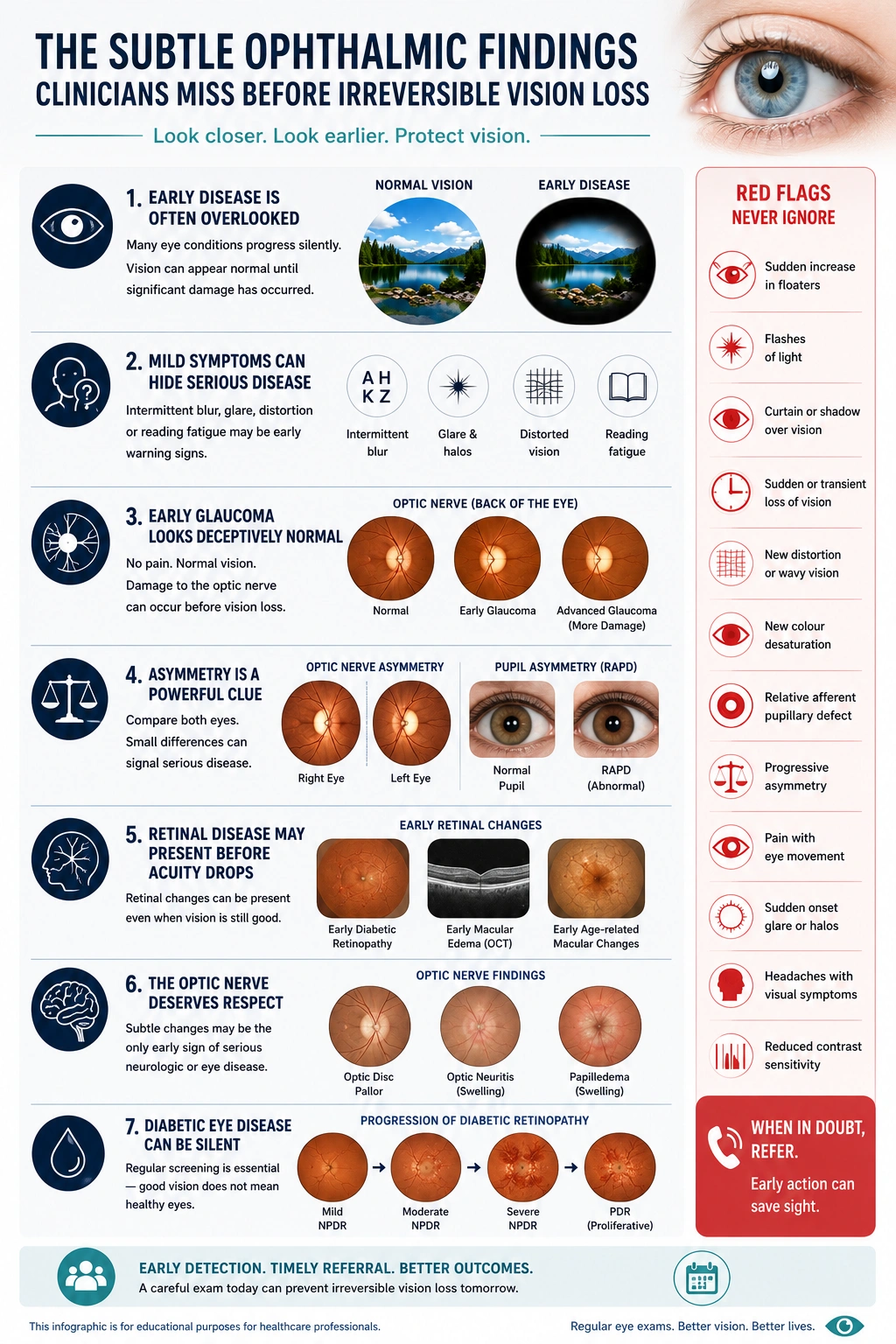
Early glaucoma often looks deceptively normal
Glaucoma remains one of the leading causes of irreversible blindness worldwide precisely because early disease is frequently missed.
In its initial stages, glaucoma may produce:
- no pain,
- no obvious redness,
- normal central visual acuity,
- and minimal patient awareness.
Subtle optic nerve asymmetry, progressive retinal nerve fibre layer thinning, or small changes in cup-to-disc ratio may represent the only early clues.
Clinicians should be particularly cautious when evaluating:
- asymmetrical cupping,
- focal neuroretinal rim thinning,
- disc haemorrhages,
- elevated intraocular pressure,
- family history,
- or unexplained visual complaints.
Importantly, a “normal” intraocular pressure does not exclude glaucoma.
Normal-tension glaucoma may progress despite pressures within statistically normal ranges, particularly in patients with vascular risk factors or compromised optic nerve perfusion.
Structural damage frequently precedes measurable functional loss.
This is why careful optic nerve assessment and appropriate imaging remain critical, even in patients who appear visually asymptomatic.
The importance of asymmetry
Asymmetry is one of the most valuable — and commonly overlooked — clinical clues in ophthalmology.
The human visual system is highly comparative. Subtle unilateral changes may become clinically significant long before bilateral dysfunction becomes obvious.
Clinicians should pay close attention to:
- asymmetric visual acuity,
- colour desaturation,
- pupil responses,
- optic nerve appearance,
- intraocular pressure,
- visual field defects,
- or symptom localisation.
A relative afferent pupillary defect (RAPD), even when subtle, may indicate:
- optic neuropathy,
- retinal disease,
- severe asymmetric glaucoma,
- or compressive pathology.
Similarly, unilateral glare complaints, asymmetric cataract density, or disproportionate visual symptoms should prompt further evaluation rather than reassurance alone.
Retinal pathology may present before acuity declines
One of the most dangerous assumptions in clinical practice is equating preserved acuity with retinal health.
Patients with early retinal disease may retain excellent central acuity while already developing:
- metamorphopsia,
- reduced contrast sensitivity,
- paracentral scotomas,
- impaired dark adaptation,
- or peripheral retinal pathology.
Subtle diabetic macular oedema, early neovascular change, epiretinal membrane formation, and age-related macular degeneration may initially produce symptoms that patients struggle to describe accurately.
Questions such as:
- “Do straight lines appear distorted?”
- “Does one eye seem dimmer?”
- “Is reading more tiring than before?”
- “Do faces appear less clear?”
- “Is night driving becoming more difficult?”
often reveal pathology that standard acuity testing alone may miss.
Dilated examination remains essential.
Without adequate retinal assessment, clinically significant pathology may remain undetected until vision loss becomes functionally obvious and structurally advanced.
The optic nerve deserves more respect
Subtle optic nerve abnormalities are among the most easily missed yet clinically important findings in medicine.
Early optic neuropathy may present with:
- mild colour vision reduction,
- brightness asymmetry,
- transient blur,
- headache,
- or vague visual complaints.
The optic disc itself may demonstrate only subtle pallor, swelling, or contour irregularity during early disease.
Clinicians should maintain a high index of suspicion in patients with:
- unexplained visual symptoms,
- pain on eye movement,
- headaches with visual change,
- transient visual obscurations,
- or asymmetric findings.
Papilledema, optic neuritis, compressive lesions, ischemic optic neuropathy, and infiltrative disease may initially appear deceptively mild.
Failure to recognise early optic nerve pathology can have profound neurologic and ophthalmic consequences.
Diabetic patients may remain asymptomatic until advanced disease
Diabetic eye disease remains one of the clearest examples of why symptom-based screening is inadequate.
Patients frequently assume that “good vision” excludes retinal disease.
In reality, clinically significant diabetic retinopathy and diabetic macular oedema may progress substantially before patients notice obvious visual decline.
Microaneurysms, intraretinal haemorrhages, venous beading, ischemia, and subtle oedema may already be present despite preserved visual acuity.
Delays in retinal screening remain a major contributor to preventable vision loss.
Clinicians managing diabetic patients should consistently reinforce that:
- retinal disease can progress silently,
- screening is preventative rather than symptom-driven,
- and early intervention dramatically improves long-term outcomes.
When glare is more than cataract
Glare is frequently attributed to ageing or cataracts without sufficient investigation.
Although cataracts are extremely common, glare symptoms may also reflect:
- ocular surface disease,
- irregular astigmatism,
- corneal pathology,
- retinal disease,
- or early macular dysfunction.
Patients describing disproportionate night driving difficulty, halos, reduced contrast, or excessive light sensitivity deserve careful assessment.
Importantly, visually significant retinal pathology may coexist with cataracts.
Assuming that cataracts alone explain visual complaints may delay the diagnosis of concurrent glaucoma, diabetic retinal disease, or macular pathology.
Clinical red flags that should never be ignored
Certain findings should consistently prompt heightened clinical concern:
- Sudden increase in floaters
- Flashes of light
- Curtain-like visual loss
- Transient monocular vision loss
- Distorted central vision
- New colour desaturation
- RAPD
- Progressive asymmetry
- Pain with eye movement
- Sudden onset glare or halos
- Persistent headaches with visual symptoms
- Unexplained reduction in contrast sensitivity
- Visual complaints despite “normal” acuity
These symptoms may represent the earliest stages of potentially vision-threatening disease.
Early recognition changes outcomes
In ophthalmology, the difference between preserved vision and irreversible blindness is often timing.
The most dangerous pathology is not always the most dramatic. Frequently, it is the subtle abnormality that progresses quietly while both patient and clinician are falsely reassured.