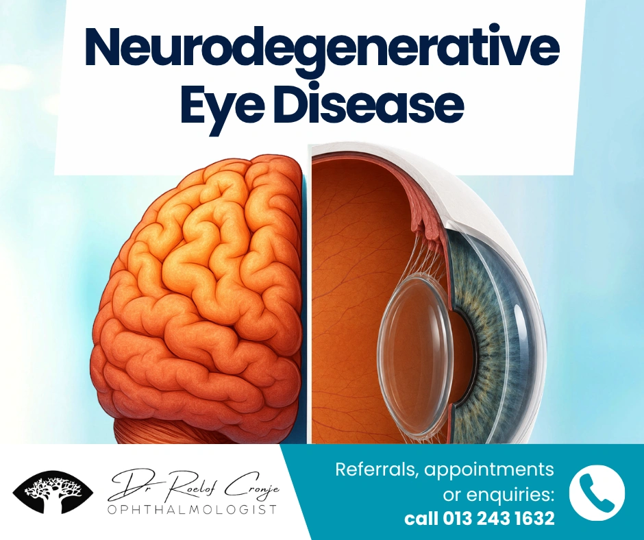
The Eyes and the Brain: An Unbreakable Connection
The eye doesn’t work in isolation. The retina and optic nerve are extensions of the brain itself, meaning that many neurological disorders also affect vision.
This link forms the basis of what we now describe as neurodegenerative eye disease — visual changes that arise from conditions such as Parkinson’s, Alzheimer’s, and Multiple Sclerosis (MS).
For optometrists and GPs, recognising subtle ocular clues can lead to earlier diagnosis, more accurate referrals, and better long-term outcomes.
Parkinson’s Disease — More Than Tremors
Parkinson’s is best known for tremor and rigidity, yet its visual effects are common and often overlooked.
Ocular Changes
- Reduced blink rate → dry eye, blepharitis, exposure irritation
- Convergence insufficiency → blurred near vision or double vision
- Slower eye movements → tracking and reading difficulties
- Reduced contrast sensitivity → problems with depth perception and mobility
Retinal Insights
OCT (Optical Coherence Tomography) frequently reveals thinning of the retinal nerve fibre layer (RNFL), reflecting dopaminergic neuron loss.
This retinal thinning parallels brain degeneration, making it a potential biomarker for neurodegenerative eye disease.
Clinical Tip
Address ocular surface discomfort, near-vision fatigue, and reduced contrast while staying alert for ocular-motor changes that may signal advancing Parkinson’s.
Alzheimer’s Disease — Seeing the Signs of Memory Loss
Alzheimer’s disease affects how patients see and process the world, even when acuity appears normal.
Ocular Changes
- Difficulty reading or recognising faces
- Reduced contrast and depth perception
- Spatial disorientation and visual confusion
Retinal Insights
Research shows retinal ganglion-cell loss and microvascular changes similar to those seen in the brain.
These findings suggest the retina could serve as a non-invasive marker of Alzheimer’s pathology.
Clinical Tip
If older patients report poor contrast, frequent missteps, or visual confusion, consider underlying cognitive decline and coordinate with their physician.
Multiple Sclerosis — When the Optic Nerve Speaks First
For many people with MS, the eye provides the first clue.
Ocular Changes
- Optic neuritis: sudden vision loss with pain on movement
- Color desaturation: reds appear pale or grey
- Double vision: due to internuclear ophthalmoplegia
- Nystagmus: rhythmic eye movements affecting focus
Retinal Insights
After optic neuritis, OCT often shows retinal nerve fibre and ganglion-cell layer thinning, even in the unaffected eye.
These measurements correlate with systemic disease activity and can help monitor progression.
Clinical Tip
In young adults with acute vision loss or color change, think optic neuritis and refer immediately for neurological assessment.
Diagnostic Tools Linking Eye and Brain
Optical Coherence Tomography (OCT)
OCT provides a quick, objective measure of neuronal health.
Thinning of the RNFL or ganglion-cell complex supports the diagnosis of neurodegenerative eye disease and helps track change over time.
Visual Field Testing
Visual field loss may reveal cortical involvement before neurological symptoms appear.
Patterns such as bitemporal or homonymous defects point to central nervous system origins rather than ocular disease alone.
When to Refer
Early collaboration between optometrists, GPs, and ophthalmologists is crucial.
Refer for specialist evaluation if you observe:
- Unexplained optic-disc pallor or asymmetry
- Color desaturation or relative afferent pupillary defect (RAPD)
- Retinal thinning on OCT without ocular cause
- Visual field loss inconsistent with glaucoma
- Persistent visual complaints in known neurological patients
Prompt referral may uncover systemic disease and prevent irreversible vision loss.
The Takeaway
The eyes don’t just reflect the soul — they reflect the brain’s health.
In many neurodegenerative disorders, the earliest signs of damage appear in the retina long before they manifest elsewhere.
By recognising the ocular manifestations of neurodegenerative eye disease, optometrists and GPs become key players in early detection, patient safety, and multidisciplinary care.
What begins in the brain can often be seen in the eye — if you know where to look.