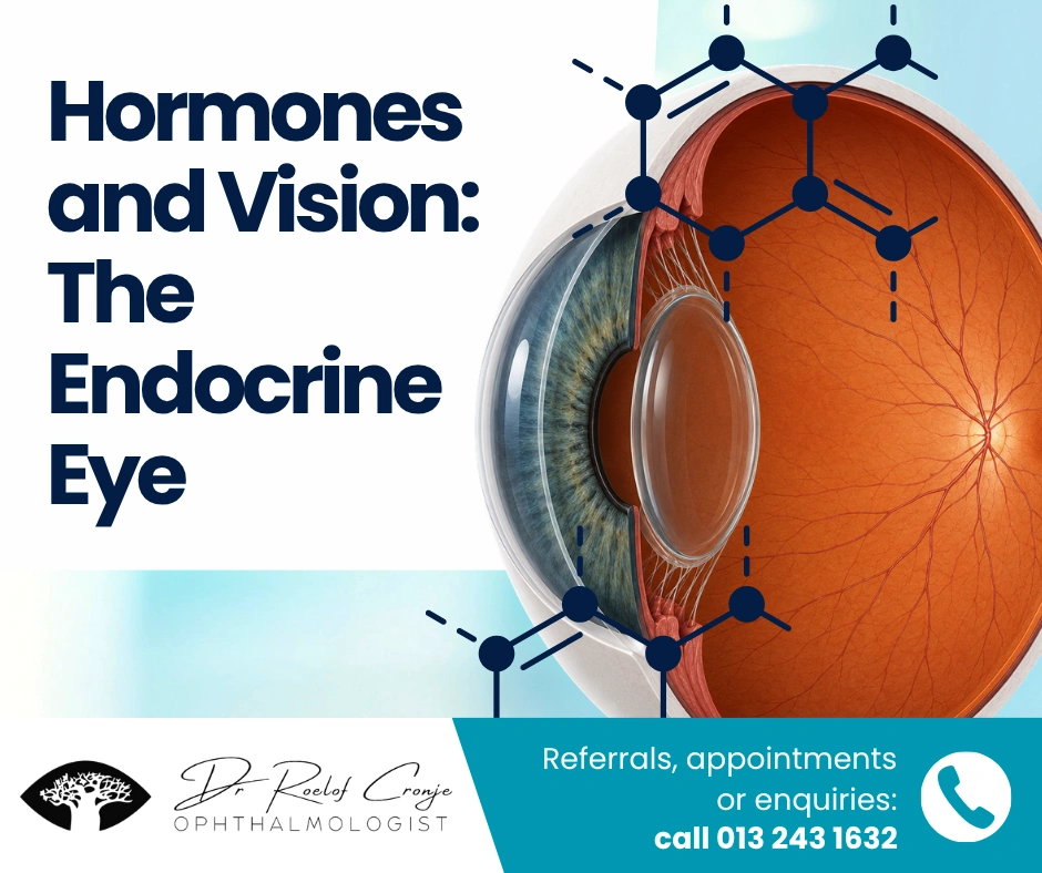
The Impact of Oestrogen, Progesterone and Hormonal Transitions on Ocular Surface, Cornea and Retina
Hormones and vision are closely linked, and endocrine disorders frequently produce measurable ocular effects. Hormonal imbalances can influence tear production, intraocular pressure, ocular surface health, and even retinal and optic nerve function.
Ocular tissues express functional receptors for oestrogen, progesterone and androgens. These receptors are present in the lacrimal gland, meibomian glands, conjunctiva, corneal epithelium, retina and optic nerve. As a result, endocrine fluctuations — whether physiological or therapeutic — can meaningfully influence visual quality, ocular comfort, refractive stability and retinal vascular integrity.
For endocrinologists and women’s health clinicians, understanding the endocrine–ocular interface enables anticipatory guidance, risk stratification and earlier multidisciplinary collaboration.
Hormonal Receptors in Ocular Tissue: A Biologically Active Environment
Sex steroid receptors are widely distributed throughout ocular structures:
- Oestrogen receptors (ER-α and ER-β) in cornea, retina, lens and lacrimal gland
- Progesterone receptors in corneal and conjunctival epithelium
- Androgen receptors in meibomian glands and lacrimal tissue
These receptors influence:
- Inflammatory signalling
- Epithelial turnover
- Lipid secretion
- Microvascular endothelial function
- Neural transmission
The ocular surface, in particular, is highly sensitive to hormonal modulation due to its reliance on delicate tear film homeostasis.
Oestrogen and the Ocular Surface
Oestrogen exerts complex effects on the tear film and ocular surface.
The tear film consists of three layers:
- Lipid (meibomian gland–derived)
- Aqueous (lacrimal gland–derived)
- Mucin (conjunctival goblet cells)
Oestrogen modulates:
- Meibomian gland lipid composition
- Goblet cell density
- Lacrimal gland inflammatory mediators
- Matrix metalloproteinase activity
Fluctuating oestrogen levels may destabilise the lipid layer, increasing tear evaporation and contributing to evaporative dry eye. This is especially relevant in peri-menopausal and postmenopausal women.
Patients often report:
- Fluctuating clarity
- Ocular fatigue
- Contact lens intolerance
- Burning or foreign body sensation
These symptoms may coincide with cyclical hormonal changes or transition periods.
Importantly, chronic ocular surface inflammation may exacerbate underlying autoimmune susceptibility.
Progesterone: Modulatory and Neuroactive Effects
Progesterone has less extensively studied but clinically relevant ocular influence.
It is believed to:
- Modulate corneal sensitivity
- Influence tear production indirectly
- Affect fluid retention within corneal stroma
Fluctuations during the luteal phase may lead to subtle corneal thickness variation, potentially affecting refractive stability. Patients may describe transient blurring or contact lens discomfort premenstrually.
While these effects are typically mild, in patients with corneal pathology or refractive surgery history, hormonal variability may amplify symptom perception.
Pregnancy: Physiological Adaptation with Ocular Consequences
Pregnancy represents a sustained high-oestrogen and progesterone state accompanied by increased plasma volume and fluid retention.
Corneal and Refractive Changes
Pregnancy may induce:
- Increased corneal thickness
- Reduced corneal sensitivity
- Temporary myopic shift
- Contact lens intolerance
These changes are generally reversible postpartum. Elective refractive procedures should be deferred during pregnancy due to refractive instability.
Intraocular Pressure
Intraocular pressure often decreases during pregnancy. Mechanisms include:
- Increased aqueous humour outflow
- Reduced episcleral venous pressure
- Hormonal effects on trabecular meshwork function
For glaucoma patients, this may temporarily reduce pressure burden, though close monitoring remains necessary.
Retinal Considerations in Pregnancy
Pregnancy may exacerbate or accelerate:
- Diabetic retinopathy
- Hypertensive retinopathy
- Central serous chorioretinopathy
In diabetic patients, rapid glycaemic normalisation may paradoxically worsen retinopathy progression. Trimester-specific retinal monitoring is recommended.
Hypertensive disorders of pregnancy may manifest with:
- Retinal arteriolar narrowing
- Cotton wool spots
- Retinal haemorrhages
- Serous retinal detachment in severe pre-eclampsia
The retina thus serves as a visible microvascular barometer during gestation.
Menopause: The Ocular Surface Shift
Menopause is one of the most significant endocrine transitions affecting the eye.
Declining oestrogen and androgen levels alter:
- Meibomian gland function
- Tear film lipid quality
- Ocular surface inflammatory balance
Postmenopausal women demonstrate higher prevalence of:
- Meibomian gland dysfunction
- Chronic evaporative dry eye
- Ocular surface inflammation
Dry eye disease in this population is not purely environmental — it reflects underlying endocrine modulation.
The condition may affect:
- Visual stability
- Quality of life
- Occupational performance
This reinforces the importance of recognising hormonal contribution to ocular discomfort rather than attributing symptoms solely to ageing.
Hormone Replacement Therapy: Nuanced Ocular Implications
The relationship between hormone replacement therapy (HRT) and ocular health remains complex.
Some studies suggest oestrogen-only therapy may increase dry eye risk, possibly through androgen suppression. Combined regimens show variable outcomes.
Considerations include:
- Duration of therapy
- Dosage
- Baseline ocular surface status
- Co-existing autoimmune disease
Additionally, retinal vascular effects of systemic hormone therapy remain under investigation. Potential modulation of endothelial function and microvascular calibre may influence retinal susceptibility in predisposed individuals.
Interdisciplinary communication is valuable when new ocular symptoms arise following HRT initiation or adjustment.
Hormones and Retinal Microvasculature
The retina is embryologically derived from neural tissue and shares microvascular architecture with cerebral circulation.
Oestrogen has been shown to influence:
- Endothelial nitric oxide synthesis
- Vascular tone
- Inflammatory cytokine expression
Declining oestrogen levels may reduce vascular protective effects, potentially contributing to increased risk of:
- Age-related macular degeneration
- Retinal vein occlusion
- Microvascular fragility
While causality remains multifactorial, the endocrine–vascular interface deserves clinical attention.
Thyroid Eye Disease: A Paradigm of Endocrine-Ocular Pathology
Thyroid-associated orbitopathy remains the most visible example of endocrine-driven ocular disease.
Pathophysiology involves:
- Autoimmune activation of orbital fibroblasts
- Glycosaminoglycan deposition
- Extraocular muscle enlargement
- Orbital congestion
Clinical features include:
- Proptosis
- Exposure keratopathy
- Diplopia
- Optic nerve compression in severe cases
Early referral is critical when visual function declines or compressive neuropathy is suspected.
The Broader Endocrine-Ocular Interface
Beyond reproductive hormones, systemic endocrine disorders with ocular implications include:
- Diabetes mellitus (microvascular retinal disease)
- Cushing’s syndrome (ocular hypertension risk)
- Hyperparathyroidism (calcific ocular deposits)
- Acromegaly (visual field compression via pituitary enlargement)
The optic nerve and retina often provide early clues to broader endocrine pathology.
Clinical Implications for Endocrinology and Women’s Health
Referral should be considered when patients experience:
- Persistent dry eye resistant to conservative therapy
- Visual distortion or metamorphopsia
- Sudden refractive shifts
- Diplopia
- New floaters or flashes
- Ocular discomfort following hormonal therapy adjustment
Routine retinal screening remains essential in:
- Diabetes
- Chronic hypertension
- Autoimmune disease
- Long-term corticosteroid use
Collaborative care improves early detection and reduces preventable visual loss.
Conclusion: The Endocrine Eye as a Clinical Opportunity
The eye is not hormonally passive. It is an endocrine-responsive organ whose surface, cornea and retina reflect systemic physiological states.
For endocrinologists and women’s health specialists, recognising hormonal influence on ocular function enhances patient counselling, anticipatory monitoring and multidisciplinary care.
The endocrine eye reminds us that vision is not isolated from systemic physiology — it is intricately integrated within it.