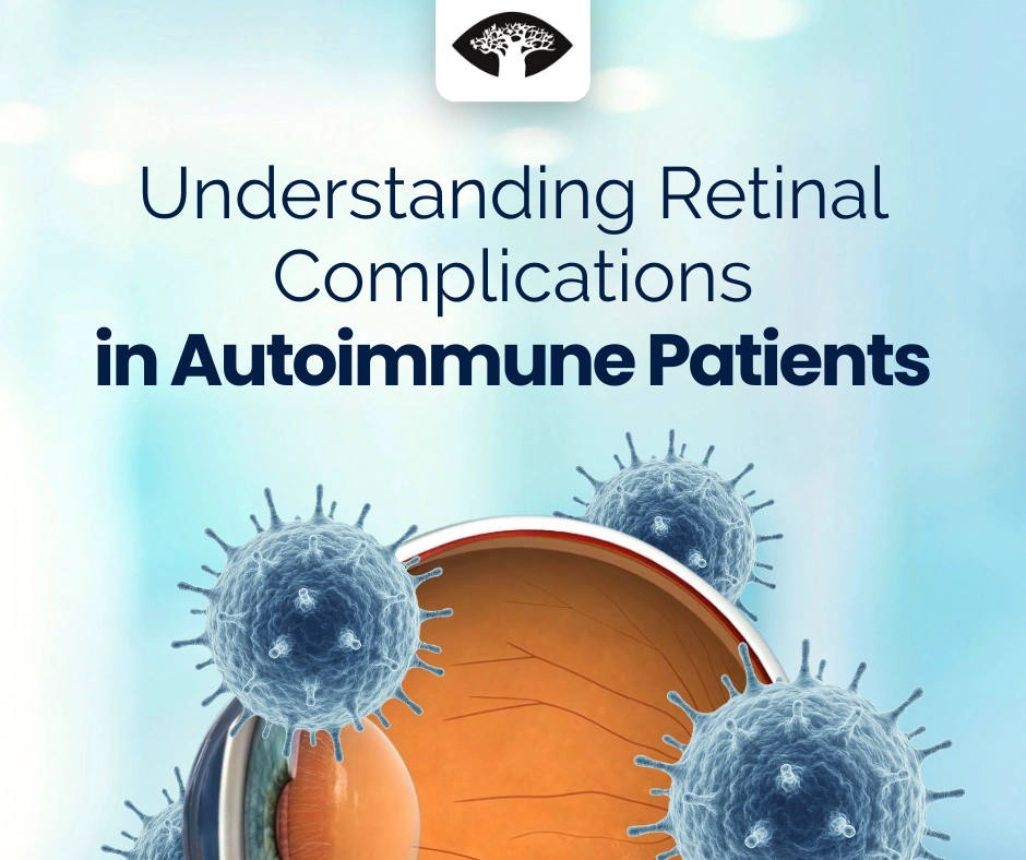
Autoimmune diseases don’t just affect joints, skin, or organs—they can also affect the eyes, especially the retina. For some patients, retinal problems may be the first sign that an autoimmune condition is active. For others, they appear later, either as complications of the disease itself or of its treatment.
For eye-care professionals, spotting these changes early can make the difference between stable vision and permanent sight loss.
How Autoimmune Diseases Affect the Retina
The immune system, when overactive or misdirected, can harm the retina in different ways:
- Direct attack – antibodies target proteins in the retina, leading to autoimmune retinopathy (AIR).
- Inflammation – vasculitis or uveitis damages retinal tissue and blood vessels
- Vascular problems – inflammation or clotting disorders such as antiphospholipid syndrome can block veins or arteries
- Medication side effects – drugs like hydroxychloroquine or steroids may damage the retina over time.
Key Retinal Complications
Autoimmune Retinopathy (AIR)
What it looks like: Progressive vision loss, flashing lights (photopsias), and blind spots. Often the retina looks normal in early stages.
Where it shows up: Reported in systemic lupus erythematosus (SLE) and paraneoplastic syndromes.
How we catch it:
- OCT: loss of outer retina.
- ERG: cone/rod dysfunction.
- FAF: patchy autofluorescence changes.
Why it matters: Can be misdiagnosed as inherited dystrophy—immunosuppression may stabilise disease.
Retinal Vasculitis
What it looks like: Cotton-wool spots, haemorrhages, vessel sheathing.
Where it shows up: Frequently seen in Behçet’s disease, SLE, and sarcoidosis.
How we catch it:
- FA highlights leakage and nonperfusion.
- OCT reveals macular oedema.
Why it matters: Untreated vasculitis can cause ischemia, neovascularisation, and severe vision loss (Palejwala, Int Ophthalmol Clin).
Macular Oedema
What it looks like: Blurred or distorted central vision, straight lines appearing wavy.
Where it shows up: Common in uveitis, juvenile idiopathic arthritis (JIA), Behçet’s disease, and sarcoidosis.
How we catch it: OCT showing cystoid macular swelling.
Why it matters: Chronic swelling damages the macula permanently if untreated.
Retinal Vein Occlusion (RVO)
What it looks like: Sudden, painless vision loss in one eye, with dilated veins and haemorrhages.
Where it shows up: Associated with antiphospholipid syndrome and SLE, due to clotting risk.
How we catch it:
- FA: blocked vessels, non-perfusion.
- OCT: macular oedema.
Why it matters: Macular oedema is the main cause of poor outcomes, but intravitreal therapy can help (Espinosa, Curr Opin Rheumatol).
Drug-Related Retinal Damage
Hydroxychloroquine Toxicity
Risk factors: Long-term use (>5 years), high cumulative dose, renal impairment.
Where it shows up: Widely prescribed in SLE and rheumatoid arthritis, with risk of toxicity rising after 5 years (Melles & Marmor, JAMA Ophthalmol).
How we catch it: OCT and FAF—early screening detects pre-symptomatic changes (Marmor et al., Ophthalmology).
Steroid-Related Complications
CSCR (central serous chorioretinopathy): strongly linked to corticosteroid use (Carvalho-Recchia, Ophthalmology).
Indirect risk: Steroids worsen diabetes, raising risk of diabetic retinopathy.
Practical Approach for Eye-Care Professionals
History: Autoimmune diagnosis, duration, treatment.
Medication review: Hydroxychloroquine, biologics, steroids.
Imaging:
- Fundus photos for documentation.
- OCT for macula and photoreceptor changes.
- FA for vascular leakage.
- ERG for suspected AIR.
Teamwork: Collaborate with rheumatologists, neurologists, and immunologists.
Why It Matters
- Retinal findings may be the first clue to autoimmune disease.
- Many complications are treatable if caught early.
- Regular monitoring helps balance systemic disease control with ocular safety.
Conclusion
Autoimmune diseases can impact the retina in multiple ways—through inflammation, vascular damage, or drug toxicity. For clinicians, using tools like OCT and FA, combined with careful history-taking and collaboration, is the key to early diagnosis and intervention.
Clinical takeaway: Always consider an autoimmune link in unexplained retinal disease, and never underestimate the value of OCT, FA, and regular screening in these patients.