What you need to know
Retinal detachment is a medical emergency because if left untreated, it can lead to permanent vision loss. When the retina detaches from the back of the eye, it is no longer able to receive oxygen and nutrients from the blood vessels that surround it. Without these vital nutrients, the retina will begin to die. If retinal detachment is not treated promptly, the retina will eventually detach completely, causing blindness.
What is retinal detachment?
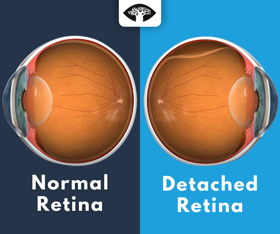
Retinal detachment is a condition in which the retina, the thin layer of tissue that lines the inside of the eye, becomes separated from the back of the eye. The retina is the innermost, light-sensitive layer of the eye and is responsible for sending visual signals to the brain. When the retina detaches, it is no longer able to function properly and vision is affected.
For a brief overview of retinal detachment watch the following informative video:
There are three types:
1. Rhegmatogenous:
A rhegmatogenous retinal detachment occurs when there is a break or tear in the retina, allowing fluid to pass through and detach the retina from the back of the eye. This type is the most common and can occur in anyone, but is more likely to happen in those with a history of eye trauma or surgery, or those who are nearsighted.
For more information on vision with a retinal tear and retinal hole watch the following informative videos:
2. Tractional:
Tractional retinal detachment occurs when abnormal growth of scar tissue on the retina pulls on the retina and causes it to detach. Scar tissue can form on the retina after an injury, surgery, or other condition that damages the eye. Treatment for tractional retinal detachment often involves surgery to repair the retina. In some cases cryotherapy (freezing) may be used to shrink the scar tissue and relieve the traction on the retina.
For a brief overview of cryotherapy watch the following:
3. Exudative:
Exudative retinal detachment is a type of retinal detachment in which fluid accumulates under the retina, causing it to lift away from the back of the eye. This is different from a rhegmatogenous, which is caused by a tear or hole in the retina. Exudative retinal detachment is usually caused by an underlying condition, such as age-related macular degeneration, that allows fluid to leak into the space under the retina.
Symptoms of retinal detachment
This condition can occur suddenly or gradually over a few days, but is considered a medical emergency. There is usually no pain associated with retinal detachment but you should all your doctor or go to the emergency room immediately if you experience any of the following symptoms:
• Sudden or gradual onset of floaters (tiny specks of debris that appear in your field of vision) and/or flashes of light in one or both eyes
• A “curtain” or “veil” appearing over your field of vision
• Cloudiness or distortion of your vision
• A sense that you are “seeing out of a tunnel”
• A decrease in your peripheral (side) vision
Risk Factors
There are several risk factors associated with retinal detachment, including:
• Myopia (nearsightedness): This is the most common risk factor. People who are nearsighted are more likely to develop this condition because their eyeballs are longer than normal. This causes the retina to be stretched thin, making it more susceptible to tearing.
• Family history: If you have a family member who has had retinal detachment, you may be at increased risk for the condition.
• Previous eye surgery: Eye surgery, such as cataract surgery, can sometimes cause retinal detachment.
• Eye injury: A severe blow to the head or face can cause retinal detachment.
• Age: It is most common in people over age 50.
Treatment Options
The most common type of treatment is surgery. There are several different types of surgery that can be used, and the best option for each individual will depend on the severity and location of the detachment.
Pneumatic retinopexy
Pneumatic retinopexy is a type of surgery that uses a gas bubble to push the retina back into place. A needle is used to inject the gas bubble into the eye, and the bubble then expands and pushes the retina back into place.
Scleral buckling
Scleral buckling is a type of surgery that involves placing a band around the outside of the eye to push the retina back into place. The band is usually made of silicone or sponge, and is placed under the eye’s surface.
Vitrectomy
Vitrectomy is a type of surgery that involves removing the vitreous gel from the eye. This gel can pull on the retina and cause it to become detached. By removing the gel, the retina is no longer being pulled on and can be pushed back into place.
Conclusion
This is a serious condition that can lead to blindness. However, if it is caught early, it can be treated successfully. There are several different treatment options available, and your doctor will work with you to determine the best course of action for your particular situation. With prompt treatment, you can often avoid serious complications and preserve your vision.