Appointments

Call Us At:
013 243 1632 or 086 166 4664

Queries

Email Us At: eye@drcronje.com

Office Hours

Mon - Fri: 8:00 AM - 5:00 PM
Sat: By Appointment Only

Categories
General Eye Health

Ocular Infectious Diseases: Prevention, Diagnosis, and Treatment

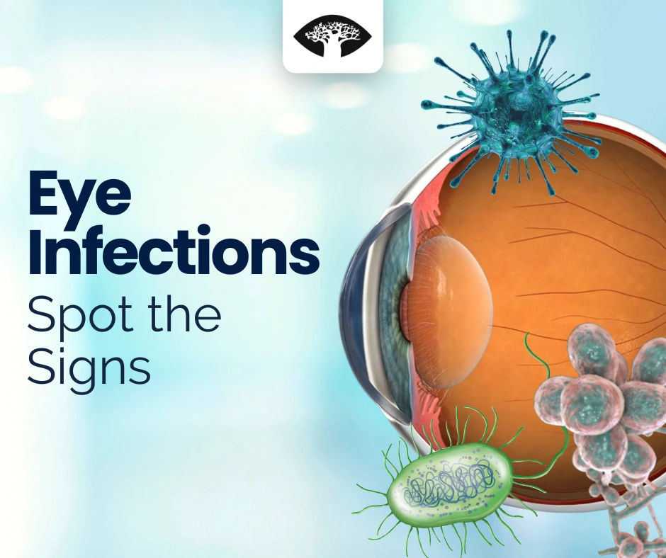
Our eyes are among the most delicate and essential organs in the body, yet they are constantly exposed to external elements that can lead to infections. Ocular infectious diseases range from mild irritations to severe conditions that can result in vision loss if left untreated. Understanding the causes, symptoms, prevention strategies, and treatment options for these infections is crucial for maintaining healthy vision.

Ocular infections arise from bacterial, viral, fungal, or parasitic sources, each presenting unique challenges in diagnosis and treatment. The impact of these infections varies from minor irritations to significant vision-threatening complications. Therefore, early detection and proper management are critical in preventing irreversible damage.

Common Ocular Infectious Diseases

Ocular infections can be caused by bacteria, viruses, fungi, or parasites, each with distinct characteristics and treatment approaches. Below are some of the most common types:

Conjunctivitis (Pink Eye)

Conjunctivitis is one of the most well-known eye infections and occurs when the thin, transparent layer covering the white part of the eye and the inner eyelids (conjunctiva) becomes inflamed.

Allergic conjunctivitis is triggered by allergens such as pollen, dust, or pet dander. Although not infectious, it causes symptoms similar to pink eye, including redness and itching. Antihistamine eye drops can provide relief.

Bacterial conjunctivitis is caused by bacteria such as Staphylococcus aureus or Streptococcus pneumoniae. It leads to redness, swelling, and thick yellow or green discharge. This type of conjunctivitis is contagious and requires antibiotic eye drops.

Viral conjunctivitis is caused by viruses like adenoviruses, the same viruses responsible for the common cold. It spreads easily and leads to watery discharge and irritation. There is no specific treatment for viral conjunctivitis, but it usually resolves on its own.

View Video

Keratitis (Corneal Infection)

The cornea is the transparent outer layer of the eye that helps focus light. Keratitis occurs when the cornea becomes infected or inflamed.

Viral keratitis, commonly caused by the Herpes Simplex Virus (HSV-1), can lead to recurrent eye infections. It causes redness, watery discharge, and in some cases, scarring of the cornea. Antiviral eye drops or oral antiviral medication can help manage the infection.

Bacterial keratitis is often seen in people who wear contact lenses and is caused by bacteria like Pseudomonas aeruginosa or Staphylococcus aureus. It can lead to severe pain, blurred vision, and eye redness. Antibiotic eye drops are required for treatment.

Fungal keratitis occurs when fungi, such as Aspergillus or Fusarium, enter the eye, usually after an injury involving plant material like a tree branch or thorn. Symptoms include eye pain, light sensitivity, and blurry vision. Antifungal eye drops or oral medication are used for treatment.

View Video

Endophthalmitis (Severe Intraocular Infection)

Endophthalmitis is a rare but severe intraocular infection that can occur following eye surgery, penetrating injuries, or bloodstream infections. It is most commonly bacterial or fungal and requires immediate medical intervention to prevent permanent vision loss.

Uveitis (Inflammation of the Uvea)

The uvea is the middle layer of the eye, which includes the iris (colored part of the eye), ciliary body (a structure that helps focus vision), and choroid (a layer filled with blood vessels). When it becomes inflamed due to infections such as toxoplasmosis (a parasite infection), syphilis, tuberculosis, or herpes viruses, it can cause pain, light sensitivity, and blurry vision. If left untreated, uveitis can lead to complications such as glaucoma (increased eye pressure) or cataracts (clouding of the lens). Anti-inflammatory and antibiotic treatments can help manage the condition.

View Video

Acanthamoeba Keratitis

Acanthamoeba keratitis is a rare but serious eye infection caused by a tiny parasite called Acanthamoeba. It is commonly found in water sources, including tap water, swimming pools, and hot tubs. People who wear contact lenses are at a higher risk if they do not properly disinfect their lenses. This infection causes extreme pain, light sensitivity, and corneal damage. Treatment is difficult and often requires specialized anti-parasitic medication.

View Video

Symptoms to Watch For

The symptoms of eye infections depend on the type of infection but commonly include:

  • Redness and swelling of the eye
  • Pain or discomfort in the eye
  • Excessive tearing or unusual eye discharge
  • Blurry vision or difficulty seeing
  • Sensitivity to light
  • The feeling that something is stuck in the eye (foreign body sensation)

If these symptoms persist, it is important to see an ophthalmologist immediately to prevent further complications.

Prevention Strategies

Preventing eye infections is possible with good hygiene and proper eye care habits. Washing hands regularly and avoiding touching the eyes can prevent bacteria and viruses from entering. People who wear contact lenses should follow proper cleaning techniques, avoid wearing them overnight, and replace them as recommended.

Sharing personal items like towels, eye makeup, and contact lenses should be avoided since infections can spread easily this way. Using protective eyewear, such as sunglasses and safety goggles, can also help shield the eyes from harmful microorganisms and environmental irritants.

Keeping the immune system strong by eating a nutritious diet, exercising, and getting enough sleep can also help the body fight infections more effectively. If an eye injury occurs, even if minor, seeking medical attention immediately can prevent secondary infections.

Diagnosis and Treatment

Ophthalmologists diagnose eye infections by performing a comprehensive eye examination. A slit-lamp examination uses a special microscope to closely examine the eye’s surface and internal structures. In some cases, an eye specialist may take a sample of eye discharge to identify the microorganism causing the infection. Fluorescein staining, where a special dye is used, can detect corneal abrasions or ulcers. PCR testing (a type of lab test) is useful in detecting viruses.

Treatment varies depending on the type of infection. Bacterial infections are treated with antibiotic eye drops, while viral infections, such as herpes keratitis, require antiviral medication. Fungal infections need antifungal eye drops or oral medication. For parasitic infections, specialized treatments may be required, often involving prolonged therapy. In severe cases, surgery, such as a corneal transplant, may be necessary to restore vision.

Conclusion

Ocular infectious diseases can range from mild conditions that resolve on their own to severe infections requiring urgent medical care. Knowing the symptoms and seeking early treatment can prevent complications. By maintaining proper eye hygiene, protecting the eyes from injury, and following medical advice, individuals can reduce their risk of developing serious eye infections.

If you notice any persistent eye discomfort, redness, or vision changes, consult an ophthalmologist immediately to ensure the best possible outcome for your eye health.

Categories
Cataracts

The Benefits of Multifocal Intraocular Lenses in Cataract Surgery

Cataract surgery is one of the most commonly performed procedures worldwide, and it has transformed the lives of millions by restoring vision lost to cataracts. A critical decision that patients face during this procedure is the choice of intraocular lenses (IOLs). Among the various options available, multifocal intraocular lenses (IOLs) stand out as a revolutionary advancement, offering a range of benefits that enhance life post-surgery.

View Video

What Are Multifocal Intraocular Lenses?

Multifocal IOLs are specialized lenses implanted during cataract surgery to replace the eye’s cloudy natural lens. Unlike traditional monofocal lenses that provide clear vision at a single distance (usually far), multifocal lenses are designed to correct vision at multiple distances, including near, intermediate, and far. This means that patients can often reduce or eliminate their dependence on glasses for everyday tasks.

View Video

Improved Vision at Multiple Distances

One of the most significant advantages of multifocal IOLs is their ability to provide clear vision at various distances. Whether you’re reading a book, working on a computer, or driving, these lenses can help you see clearly without the need for multiple pairs of glasses or bifocals. This comprehensive range of vision is a game-changer for many patients, as it restores their ability to engage in daily activities seamlessly.

Reduced Dependence on Glasses

For individuals who have relied on glasses for most of their lives, multifocal IOLs can be life-changing. Many patients report a dramatic reduction in their dependence on glasses for activities such as reading or watching television. This convenience improves quality of life and enhances confidence in social and professional settings.

Enhanced Quality of Life

Multifocal IOLs often lead to an improved quality of life. Patients regain the freedom to perform various tasks without constant interruptions caused by switching glasses. From enjoying outdoor activities to working on detailed crafts or hobbies, multifocal lenses make these experiences more accessible and enjoyable.

Advanced Technology for Modern Lifestyles

The design of multifocal IOLs incorporates cutting-edge optical technology. Some lenses use diffractive designs to distribute light effectively across different focal points, ensuring clear vision in varying lighting conditions. This adaptability is particularly beneficial for modern lifestyles, where tasks often require frequent shifts between near and distant vision.

Considerations When Choosing Multifocal IOLs

While the benefits of multifocal lenses are compelling, it is essential to consider individual needs and expectations. Factors such as lifestyle, occupation, and the presence of other eye conditions can influence the suitability of multifocal IOLs. Consulting with an experienced ophthalmologist is crucial to making an informed decision that aligns with your visual goals.

Are Multifocal IOLs Right for You?

Not every patient is a candidate for multifocal IOLs. Certain conditions, such as severe astigmatism or specific corneal abnormalities, may require alternative options. Additionally, while multifocal lenses can significantly reduce dependence on glasses, some patients may still need reading glasses for very fine print or low-light situations.

View Video

Conclusion

Multifocal intraocular lenses represent a remarkable advancement in cataract surgery, offering patients the opportunity to regain clear vision at multiple distances and reduce their dependence on glasses. By understanding the benefits and considerations of these lenses, patients can make informed choices that align with their lifestyle and vision goals.

If you are considering cataract surgery, consult with your ophthalmologist to determine whether multifocal IOLs are the right option for you. With the right lens choice, you can look forward to a future of enhanced vision and a renewed sense of independence.

Categories
Uncategorised

The Role of Genetics in Eye Health: Insights into Family History and Vision Care

Your family’s health history can reveal a lot about your potential risk for developing certain medical conditions, and your eyes are no exception. Many eye diseases have a genetic component, meaning that a predisposition to develop these conditions can be inherited from your parents or grandparents. Understanding these hereditary risks can empower you to take proactive steps toward protecting your vision and maintaining your quality of life.

View Video

Common Hereditary Eye Conditions

  1. Macular Degeneration
    • What It Is: Age-related macular degeneration (AMD) is a leading cause of vision loss among people over 50. It affects the macula, the central part of the retina responsible for sharp central vision.
    • Genetic Link: Certain genetic variations, such as mutations in the CFH and ARMS2 genes, are associated with an increased risk of AMD.
    • Signs to Watch For: Blurred central vision, difficulty recognizing faces, or straight lines appearing wavy.
View Video
  1. Glaucoma
    • What It Is: Glaucoma damages the optic nerve, which sends visual information from your eyes to your brain. It’s often caused by high pressure inside the eye.
    • Family Link: If someone in your family has glaucoma, you’re more likely to get it too. Early detection and treatment can prevent significant vision loss.
    • Warning Signs: Losing side vision, tunnel vision, or eye pain in severe cases. However, most forms of glaucoma show no symptoms until advanced stages, making regular screenings essential.
View Video
  1. Retinitis Pigmentosa
    • What It Is: This is a rare condition that slowly damages the retina, causing vision loss over time. It’s one of the most well-known hereditary eye diseases.
    • Family Link: It’s usually inherited, and there are different patterns of inheritance based on your family’s genetics. Understanding your family’s history can guide early interventions.
    • Warning Signs: Trouble seeing at night, losing side vision, or struggling in dim lighting. Genetic counseling can help determine risk levels for family members.
View Video
  1. Keratoconus
    • What It Is: A condition where the cornea thins and bulges into a cone-like shape, causing distorted vision.
    • Genetic Link: Family history is a significant risk factor, and researchers are identifying specific genetic markers associated with keratoconus.
    • Signs to Watch For: Blurry vision, light sensitivity, and frequent changes in eyeglass prescriptions.
View Video

How to Protect Your Vision

If you know that eye problems run in your family, there are proactive steps you can take to reduce your risk and detect issues early:

  1. Get Regular Eye Exams
    • Seeing your eye doctor regularly can help catch problems early, even before you notice symptoms. Inform your doctor about your family’s eye health history to ensure personalized care.
    • Eye pressure tests, retinal scans, and genetic testing may be recommended for higher-risk patients.
View Video
  1. Adopt a Healthy Lifestyle
    • Balanced Diet: Include foods rich in omega-3 fatty acids, lutein, and antioxidants. Leafy greens, fish, eggs, nuts, and citrus fruits support eye health and reduce inflammation.
    • Exercise: Regular physical activity improves blood flow to your eyes and lowers the risk of glaucoma and other vascular-related eye issues.
    • Quit Smoking: Smoking accelerates eye diseases like macular degeneration and increases the risk of cataracts and dry eyes.
  2. Protect Your Eyes
    • UV Protection: Wear sunglasses that block 100% of UV rays. Overexposure to sunlight can contribute to cataracts and other eye conditions.
    • Safety Eyewear: Use protective eyewear during sports, home improvement projects, or activities that pose risks to your eyes.
  3. Monitor Vision Changes
    • Keep a journal of any changes in your vision, such as trouble focusing, blurry spots, or night blindness. Promptly report these to your eye doctor for early evaluation.
  4. Consider Genetic Counseling and Testing
    • Genetic counseling can help you understand your inherited risks for specific eye conditions. Testing may provide insights into preventive strategies and guide your care plan.
View Video

Taking Control of Your Eye Health

Your genetic makeup is just one piece of the puzzle when it comes to your eye health. By combining regular check-ups, a healthy lifestyle, and the latest medical advancements, you can greatly reduce the impact of hereditary conditions. Being proactive ensures that your vision remains as clear and healthy as possible for years to come.

Categories
Retinal Conditions

Understanding and Managing Age-Related Macular Degeneration (AMD)

Age-Related Macular Degeneration (AMD) is a leading cause of vision loss among individuals over 50, affecting the macula—the central part Understanding and Managing Age-Related Macular Degeneration (AMD)

What Is AMD?

AMD is a condition that affects the macula, the central part of the retina at the back of the eye. The macula is responsible for sharp, detailed central vision, which is essential for tasks like reading, driving, and recognizing faces. When the macula deteriorates, it leads to vision loss in the center of the visual field, while peripheral (side) vision remains intact.

View Video

Prevalence of AMD in South Africa

In South Africa, the prevalence of AMD is a growing concern. A study conducted in Durban found that among older adults living in low-income old age homes, 3.88% had AMD, making it the third leading cause of visual impairment in this population.

SciELO

Causes of AMD

The exact cause of AMD remains unclear, but it is believed to involve a combination of genetic, environmental, and lifestyle factors. The macula contains light-sensitive cells that can deteriorate over time, leading to the symptoms associated with this condition.

Dr Roelof Cronje

Symptoms of AMD

Early stages of AMD may present no symptoms. As the condition progresses, individuals might experience:

  • Blurred or distorted central vision
  • Difficulty seeing in low-light conditions
  • Faded or washed-out colors
  • A dark or empty area in the center of vision

These symptoms can interfere with tasks like reading, driving, and recognizing faces.

Types of AMD

AMD is categorized into two main types:

  1. Dry AMD (Non-Neovascular):
    The more common form, accounting for about 85-90% of cases. It progresses slowly and features the presence of drusen (small yellow deposits under the retina) and thinning of the macular tissue. Dr Roelof Cronje
View Video
  1. Wet AMD (Neovascular):
    Less common but more severe, wet AMD involves the growth of abnormal blood vessels under the retina that can leak fluid and blood, leading to rapid vision loss. Dr Roelof Cronje
View Video

Risk Factors

Several factors can increase the risk of developing AMD:

  • Age: The risk increases with age, particularly after 50.
  • Family History: A genetic predisposition can elevate risk.
View Video
  • Smoking: Smoking significantly increases the risk of AMD.
  • Obesity: Higher body mass index is associated with increased risk.
  • Sunlight Exposure: Prolonged exposure to UV light may contribute to AMD development.
View Video

Diagnosis

AMD is diagnosed through a comprehensive eye examination, which may include:

  • Visual Acuity Test: Measures the sharpness of vision.
  • Dilated Eye Exam: Allows the doctor to view the retina and macula.
  • Amsler Grid Test: Detects visual distortions.
View Video
  • Fluorescein Angiography: Uses dye to highlight blood vessels in the eye.
View Video

Treatment Options

While there is no cure for AMD, several treatments can help manage the condition and slow its progression:

  • Anti-VEGF Injections: Medications like Avastin and Lucentis are injected into the eye to inhibit the growth of abnormal blood vessels in wet AMD. Dr Roelof Cronje
View Video
  • Photodynamic Therapy: A light-activated drug is used to target and destroy abnormal blood vessels in wet AMD. Dr Roelof Cronje
  • Nutritional Supplements: The Age-Related Eye Disease Study (AREDS) found that a specific combination of vitamins and minerals can reduce the risk of progression in intermediate or advanced AMD. Dr Roelof Cronje
View Video

Lifestyle Modifications

Adopting certain lifestyle changes can help manage AMD:

  • Diet: Consuming a diet rich in leafy greens, fish, and nuts can support eye health.
  • Exercise: Regular physical activity may reduce the risk of AMD progression.
  • Smoking Cessation: Quitting smoking can significantly lower the risk.
  • UV Protection: Wearing sunglasses that block UV light can protect the eyes.

Conclusion

Age-Related Macular Degeneration (AMD) is a leading cause of vision loss among individuals over 50, affecting the macula—the central part of the retina responsible for sharp, detailed vision. This progressive condition can significantly impact daily activities, including reading, driving, and recognizing faces.

Categories
Retinal Conditions

How Blue Light Exposure Affects Your Macula

In today’s digital age, our eyes are exposed to more screens than ever before—phones, computers, tablets, and TVs dominate our daily routines. One of the key concerns that has emerged is the impact of blue light on eye health, particularly on the macula, a vital part of the retina responsible for sharp central vision. In this article, we’ll explore how blue light affects your macula, its potential risks, and how you can protect your vision.

What Is Blue Light?

Blue light is a high-energy, short-wavelength light that is naturally emitted by the sun. However, artificial sources such as LED screens, fluorescent lighting, and digital devices also produce significant amounts of blue light. While blue light exposure during the day can help regulate our sleep-wake cycle, prolonged exposure—especially at night—has raised concerns about its effects on eye health.

View Video

Understanding the Macula’s Role in Vision

The macula is a small but crucial part of the retina located at the back of the eye. It is responsible for detailed central vision, which allows us to perform tasks like reading, driving, and recognizing faces. Because of its high concentration of photoreceptor cells, the macula is particularly vulnerable to damage from light exposure, including blue light.

View Video

How Blue Light Affects the Macula

  1. Oxidative Stress
    Blue light penetrates deep into the eye, reaching the macula. Its high energy can generate oxidative stress, which damages the photoreceptor cells in the macula over time. This can increase the risk of conditions like macular degeneration.
  2. Disruption of Protective Pigments
    The macula contains pigments such as lutein and zeaxanthin that act as natural filters, shielding it from harmful light. Prolonged blue light exposure can deplete these pigments, reducing their protective ability and leaving the macula more vulnerable to damage.
  3. Potential Acceleration of Age-Related Macular Degeneration (AMD)
    Studies suggest that prolonged blue light exposure may contribute to the development or worsening of age-related macular degeneration, a leading cause of vision loss in older adults. While more research is needed, this potential link underscores the importance of preventative measures.
View Video

Symptoms of Blue Light Strain

If you spend long hours in front of screens, you may experience symptoms such as:

  • Eye fatigue and discomfort
  • Blurred vision
  • Dry eyes
  • Difficulty focusing
  • Headaches, particularly after prolonged device use

While these symptoms are often temporary, chronic exposure without protective measures may lead to more significant long-term issues.

Protecting Your Macula from Blue Light

Fortunately, there are several steps you can take to minimize the impact of blue light on your macula and overall eye health:

  1. Use Blue Light-Blocking Glasses
    These specially designed glasses filter out harmful blue light, reducing the strain on your eyes during screen use.
  2. Apply the 20-20-20 Rule
    Every 20 minutes, take a 20-second break and look at something 20 feet away. This practice helps reduce eye strain from prolonged screen time.
  3. Adjust Your Screen Settings
    Many devices have a “night mode” or blue light filter setting that reduces the intensity of blue light emitted, particularly during evening hours.
  4. Increase Lutein and Zeaxanthin Intake
    Consuming leafy greens like spinach, kale, and other lutein- and zeaxanthin-rich foods can strengthen the macula’s natural defenses.
  5. Limit Screen Time
    Where possible, take regular breaks from digital devices, especially before bedtime. This will not only help your eyes but also improve your sleep quality.
  6. Wear UV-Blocking Sunglasses
    Although primarily aimed at outdoor light, sunglasses that block UV and blue light can offer additional protection.
View Video

When to Seek Professional Help

If you experience persistent symptoms of eye strain, blurry vision, or discomfort, it’s essential to consult an eye care professional. Regular comprehensive eye exams can detect early signs of macular damage and other vision problems.

Conclusion

While blue light is an inevitable part of modern life, taking steps to protect your macula can help safeguard your vision for years to come. Simple changes like wearing blue light-blocking glasses, following the 20-20-20 rule, and eating a diet rich in protective nutrients can make a significant difference. By prioritizing eye health today, you can reduce the risk of long-term damage and continue enjoying clear, vibrant vision.

Appointments

Call Us At:
013 243 1632 or 086 166 4664

Queries

Email Us At: eye@drcronje.com

Office Hours

Mon - Fri: 8:00 AM - 5:00 PM
Sat: By Appointment Only

The information on this website is for educational purposes only and is not a substitute for professional medical advice, diagnosis, or treatment. Please consult Dr. Cronje for specific eye health concerns.

Request to Schedule Appointment

Please provide two possible dates that suit you, we will revert to you to discuss availability according to Dr. Cronje’s schedule.

All information provided below is private and protected by the POPI act

Open chat
Scan the code
Have a Question? Let’s Chat!
I’m here to help with any questions you have about eye care or treatments.