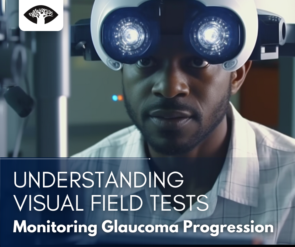
Glaucoma is a chronic eye condition that can lead to irreversible vision loss if left untreated. It affects the optic nerve, which is responsible for transmitting visual information from the eye to the brain. As the disease progresses, the optic nerve fibers become damaged, resulting in visual field loss. Visual field tests are crucial tools used by ophthalmologists to monitor glaucoma progression and assess the effectiveness of treatment.
What is a Visual Field Test?
A visual field test, also known as perimetry, is a diagnostic procedure that measures a person’s entire field of vision, including their central and peripheral vision. The test provides valuable information about the extent and pattern of visual field loss, which is a key indicator of glaucoma progression. There are various types of visual field tests, including automated perimetry, frequency doubling technology, and more.
Static perimetry is the most commonly used type of visual field test. It involves presenting a series of light stimuli at different locations in the visual field, and the patient indicates when they see the light by pressing a button. Kinetic perimetry, on the other hand, uses moving stimuli to map the boundaries of the visual field.
How is a Visual Field Test Performed?
Before the test, the patient’s eyes are typically dilated to ensure a clear view of the retina. The patient sits in a dimly lit room and places their chin on a chin rest, while one eye is covered to assess each eye separately. The technician operates the equipment and guides the patient through the test.
During the test, the patient focuses on a central fixation point and responds whenever they see a stimulus. The stimuli are presented at varying intensities and locations within the visual field. The patient’s responses are recorded, and the test continues until a comprehensive assessment of the visual field is obtained.
Different perimetry strategies may be used, depending on the purpose of the test. Threshold strategies involve presenting stimuli at different intensities to determine the minimum level of brightness the patient can perceive. Suprathreshold strategies use stimuli that are presented above the patient’s threshold, making the test faster but less sensitive to subtle visual field defects.
Interpreting Visual Field Test Results
Visual field test results are typically provided in a visual field report, which includes several components. These components may include a visual field map, showing the areas of normal and abnormal vision, and numerical indices that provide quantitative measurements of visual field loss.
The location of visual field defects is significant in understanding glaucoma progression. Common types of defects include paracentral defects, which affect the central part of the visual field, and arcuate defects, which manifest as a loss of vision in a curved or arc-like shape.
Different classification systems are used to grade visual field defects and assess the severity of glaucoma. Examples include the Glaucoma Hemifield Test, which compares the upper and lower halves of the visual field, and the Mean Deviation and Pattern Standard Deviation, which provide information about the overall deviation from normal and the pattern of visual field loss, respectively.
Factors that Can Affect Visual Field Test Results
Several factors can influence the reliability and accuracy of visual field test results. Patient fatigue is a common concern, as prolonged testing sessions may lead to reduced attention and response accuracy. Learning effects can also impact results, as patients may become more familiar with the test and perform better over time.
Other factors that can affect visual field test results include media opacities, such as cataracts, which can scatter light and distort the test stimuli. Eye movements and fixation losses during the test can introduce errors, leading to false positives (incorrectly indicating the presence of a stimulus) or false negatives (omitting the presence of a stimulus). Proper patient instruction and technician guidance are essential to minimize these errors.
The Role of Visual Field Tests in Glaucoma Management
Visual field tests play a crucial role in monitoring glaucoma progression and assessing the effectiveness of treatment. They provide valuable information to ophthalmologists, helping them make informed decisions regarding the management of the disease. By detecting visual field defects, visual field tests can indicate the need for treatment adjustments or interventions to prevent further vision loss.
The frequency of visual field testing in glaucoma management varies based on individual patient factors, such as the severity of the disease and the stability of visual field defects. In the early stages of glaucoma, visual field tests may be performed every 6 to 12 months to establish a baseline and track disease progression. As the disease advances, more frequent testing, such as every 3 to 6 months, may be necessary to closely monitor changes and guide treatment decisions.
Visual field tests are integrated with other clinical information, including intraocular pressure measurements, optic nerve assessments, and imaging tests, to provide a comprehensive evaluation of the patient’s condition. The collective data help ophthalmologists determine the optimal treatment approach and evaluate the effectiveness of interventions over time.
Conclusion
Visual field tests are invaluable tools in the management of glaucoma. By assessing the extent and pattern of visual field loss, these tests provide essential information for monitoring disease progression and guiding treatment decisions. Patients with glaucoma should work closely with their ophthalmologist to understand their visual field test results and their implications for their overall glaucoma management.
Regular visual field testing, in combination with other clinical assessments, helps ensure early detection of glaucoma progression and appropriate intervention to preserve vision. By staying proactive and engaged in their eye care, patients can actively contribute to maintaining their visual health and quality of life.


