Appointments

Call Us At:
013 243 1632 or 086 166 4664

Queries

Email Us At: eye@drcronje.com

Office Hours

Mon - Fri: 8:00 AM - 5:00 PM
Sat: By Appointment Only

Categories
Retinal Conditions

Everything You Need To Know About Retinal Detachment
What you need to know

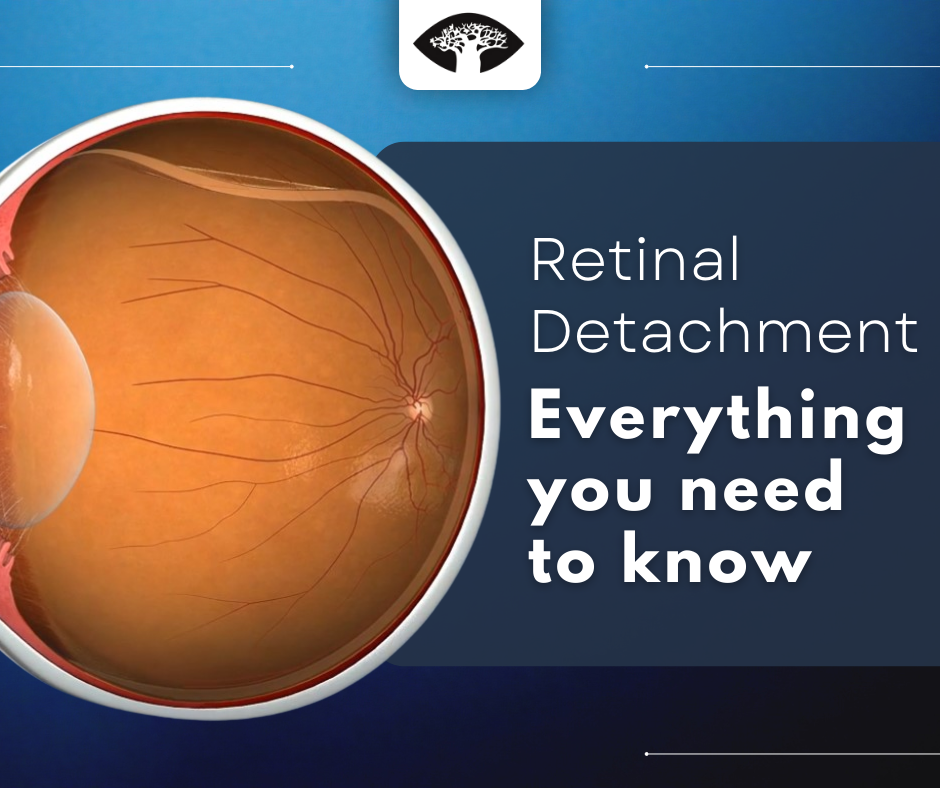
Retinal detachment is a medical emergency because if left untreated, it can lead to permanent vision loss. When the retina detaches from the back of the eye, it is no longer able to receive oxygen and nutrients from the blood vessels that surround it. Without these vital nutrients, the retina will begin to die. If retinal detachment is not treated promptly, the retina will eventually detach completely, causing blindness.

What is retinal detachment?

Retinal detachment is a condition in which the retina, the thin layer of tissue that lines the inside of the eye, becomes separated from the back of the eye. The retina is the innermost, light-sensitive layer of the eye and is responsible for sending visual signals to the brain. When the retina detaches, it is no longer able to function properly and vision is affected.

Infographic showing the difference between a normal eye and an eye with retinal detachment

For a brief overview of retinal detachment watch the following informative video:

View Video

There are three types:

1. Rhegmatogenous:

A rhegmatogenous retinal detachment occurs when there is a break or tear in the retina, allowing fluid to pass through and detach the retina from the back of the eye. This type is the most common and can occur in anyone, but is more likely to happen in those with a history of eye trauma or surgery, or those who are nearsighted.

For more information on vision with a retinal tear and retinal hole watch the following informative videos:
2. Tractional:

Tractional retinal detachment occurs when abnormal growth of scar tissue on the retina pulls on the retina and causes it to detach. Scar tissue can form on the retina after an injury, surgery, or other condition that damages the eye. Treatment for tractional retinal detachment often involves surgery to repair the retina. In some cases cryotherapy (freezing) may be used to shrink the scar tissue and relieve the traction on the retina.

For a brief overview of cryotherapy watch the following:
View Video
3. Exudative:

Exudative retinal detachment is a type of retinal detachment in which fluid accumulates under the retina, causing it to lift away from the back of the eye. This is different from a rhegmatogenous, which is caused by a tear or hole in the retina. Exudative retinal detachment is usually caused by an underlying condition, such as age-related macular degeneration, that allows fluid to leak into the space under the retina.

Symptoms of retinal detachment

This condition can occur suddenly or gradually over a few days, but is considered a medical emergency. There is usually no pain associated with retinal detachment but you should all your doctor or go to the emergency room immediately if you experience any of the following symptoms:

• Sudden or gradual onset of floaters (tiny specks of debris that appear in your field of vision) and/or flashes of light in one or both eyes

• A “curtain” or “veil” appearing over your field of vision

• Cloudiness or distortion of your vision

• A sense that you are “seeing out of a tunnel”

• A decrease in your peripheral (side) vision

Risk Factors

There are several risk factors associated with retinal detachment, including:

Myopia (nearsightedness): This is the most common risk factor. People who are nearsighted are more likely to develop this condition because their eyeballs are longer than normal. This causes the retina to be stretched thin, making it more susceptible to tearing.

Family history: If you have a family member who has had retinal detachment, you may be at increased risk for the condition.

Previous eye surgery: Eye surgery, such as cataract surgery, can sometimes cause retinal detachment.

Eye injury: A severe blow to the head or face can cause retinal detachment.

Age: It is most common in people over age 50.

Treatment Options

The most common type of treatment is surgery. There are several different types of surgery that can be used, and the best option for each individual will depend on the severity and location of the detachment.

Pneumatic retinopexy

Pneumatic retinopexy is a type of surgery that uses a gas bubble to push the retina back into place. A needle is used to inject the gas bubble into the eye, and the bubble then expands and pushes the retina back into place.

View Video

Scleral buckling

Scleral buckling is a type of surgery that involves placing a band around the outside of the eye to push the retina back into place. The band is usually made of silicone or sponge, and is placed under the eye’s surface.

View Video

Vitrectomy

Vitrectomy is a type of surgery that involves removing the vitreous gel from the eye. This gel can pull on the retina and cause it to become detached. By removing the gel, the retina is no longer being pulled on and can be pushed back into place.

View Video

Conclusion

This is a serious condition that can lead to blindness. However, if it is caught early, it can be treated successfully. There are several different treatment options available, and your doctor will work with you to determine the best course of action for your particular situation. With prompt treatment, you can often avoid serious complications and preserve your vision.

Categories
General Eye Health

Food For Thought: How What You Eat Affects Your Eye Health
Your Diet And Eye Health

Your diet has a big impact on your eye health. The foods you eat can affect your vision in both the short and long term. A diet that is high in fat and sugar can lead to problems such as obesity and diabetes, which can in turn lead to serious eye conditions such as glaucoma and cataracts. Conversely, a diet that is rich in antioxidants and nutrients can help to protect your eyes from age-related damage and disease.

A healthy diet can help you maintain a healthy weight.

There are a number of eye problems that people who are overweight or obese are at an increased risk for. These include glaucoma, diabetic retinopathy, and age-related macular degeneration. Each of these conditions can lead to vision loss or blindness if left untreated.

For tips on portion control for healthy weight management watch this informative video:

View Video

Eating a diet rich in fruits and vegetables can help protect your eyes from age-related vision problems.

Fruits and vegetables contain antioxidants that help to keep your eyes healthy. Some antioxidants found in fruits and vegetables that are beneficial for eye health include vitamin C, beta-carotene, and flavonoids. These nutrients help to protect the eyes from damage caused by free radicals, which can lead to conditions such as macular degeneration and cataracts. Eating a diet rich in fruits and vegetables is one of the best ways to ensure that your eyes stay healthy and to reduce your risk of developing these conditions.

Eating a diet high in omega-3 fatty acids can help to protect your eyes from dry eye syndrome and age-related macular degeneration.

Omega-3 fatty acids are a type of unsaturated fat that is found in oily fish such as salmon, mackerel, and sardines, as well as in flaxseeds, chia seeds, and walnuts. They are essential for maintaining the health of cell membranes, and they have anti-inflammatory properties that can help to reduce the risk of a number of conditions, including dry eye syndrome and age-related macular degeneration. If you are concerned about your eye health, you should speak to your doctor or a registered dietitian about incorporating more omega-3 fatty acids into your diet.

To learn more about the benefits of omega-3 watch this informative video:

View Video

Eating a diet high in sugar can increase your risk for diabetic retinopathy.

A diet high in sugar can lead to diabetes, which in turn can cause diabetic retinopathy. Diabetic retinopathy is a serious medical condition in which the blood vessels in the retina are damaged, potentially leading to blindness. If you have diabetes, it is important to control your blood sugar levels to help prevent this complication.

Eating a diet high in salt can increase your risk for glaucoma.

Salt is an important part of our diet, but eating too much salt can increase our risk for developing high blood pressure, which is a major risk factor for developing glaucoma. People with glaucoma have an increased pressure in their eyes, which can damage the optic nerve and lead to vision loss. By following a healthy diet, you can help to protect your eyesight and maintain good vision for years to come.

Other nutrients you can consume to help protect your eyes from disease and keep them healthy include:

Vitamin A: Vitamin A helps to produce tears and is important for night vision. It can be found in foods like carrots, sweet potatoes, and spinach.

Vitamin C: Vitamin C helps to protect the cornea, which is the clear, outer layer of the eye. It can be found in foods like oranges, strawberries, and kale.

Vitamin E: Vitamin E helps to protect the cells in the retina, which is the back part of the eye that senses light. It can be found in foods like almonds, sunflower seeds, and avocados.

Categories
Diabetic Retinopathy

6 Tips To Prevent The Progression Of Diabetic Retinopathy
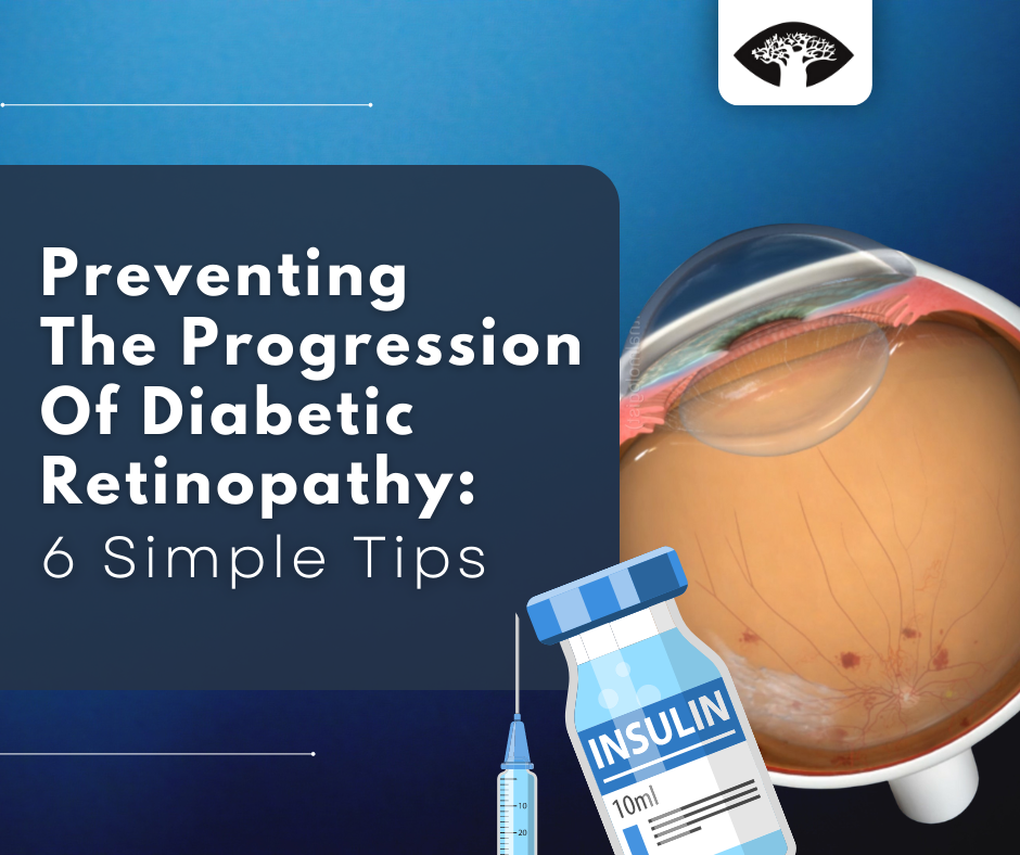
If you have diabetes, you know that it is important to keep your blood sugar levels under control to prevent complications. But did you know that you also need to take care of your eyes?

Diabetic retinopathy is a leading cause of blindness in adults, and it can occur even if your blood sugar is well-controlled. Here are five tips from an ophthalmologist to help you prevent the progression of diabetic retinopathy:

Get your eyes checked regularly.


Diabetic retinopathy is a condition that affects the blood vessels in the retina, the light-sensitive tissue at the back of the eye. The condition is a leading cause of blindness in adults. People with diabetes are at risk for developing diabetic retinopathy. The condition often has no early symptoms, so it is important to have a comprehensive dilated eye exam at least once a year. Your ophthalmologist can detect the early signs of the disease and start treatment to prevent vision loss.

To learn more about the importance of routine eye exams for diabetics watch the following informative video:

View Video

Keep your blood sugar levels under control.

Diabetes prevents your body from using and storing sugar properly, which can cause excessive amounts of sugar to remain in the bloodstream. The resulting high glucose levels can damage blood vessels throughout the body, including those in the eyes.

Maintaining good blood sugar control is the most important thing you can do to prevent diabetic retinopathy. Work with your doctor to keep your blood sugar levels as close to normal as possible.

If you have high blood sugar for an extended period of time, it can change the shape of your eye’s lens, making your vision blurry. If left uncontrolled, it can result in blindness.

Keep your blood pressure under control.

High blood pressure can damage the blood vessels in your eyes, so it is important to keep it under control. Work with your doctor to keep your blood pressure at a healthy level.

If you have high blood pressure, it is important to take measures to lower it. This can include taking medications and reducing stress levels. Uncontrolled blood pressure can result in the bursting of thin blood vessels in your eyes.

To learn more about monitoring your blood pressure watch this helpful video:

View Video

Quit smoking.

All types of tobacco contain cyanide, a poisonous substance that can damage cells in the eyes. Additionally, tobacco use can interfere with proper blood circulation and oxygenation. Smoking damages the blood vessels in your eyes and makes diabetic retinopathy worse. If you smoke, quit. It’s the best thing you can do for your eyes.

Eat a healthy diet.

Eating a diet that is rich in fruits, vegetables, and whole grains can help protect your eyes from diabetic retinopathy. So eat healthy and take care of your eyes!

Appointments

Call Us At:
013 243 1632 or 086 166 4664

Queries

Email Us At: eye@drcronje.com

Office Hours

Mon - Fri: 8:00 AM - 5:00 PM
Sat: By Appointment Only

The information on this website is for educational purposes only and is not a substitute for professional medical advice, diagnosis, or treatment. Please consult Dr. Cronje for specific eye health concerns.

Request to Schedule Appointment

Please provide two possible dates that suit you, we will revert to you to discuss availability according to Dr. Cronje’s schedule.

All information provided below is private and protected by the POPI act

Open chat
Scan the code
Have a Question? Let’s Chat!
I’m here to help with any questions you have about eye care or treatments.